LCMS & MALDI Newsletter - Mass Spectrometry Imaging Issue
Check out the latest interviews, and find out information on the state of the art instrument and software updates currently available from Shimadzu!
Contents
- Introduction
- Dr Ruth Gordillo, Southwestern Metabolic Phenotyping Core, University of Texas
Enhancing Analytical Capabilities in Biomedical Research – the Shimadzu iMScope QT enables spatial resolution and assists to uncover subtle spatially localized metabolite differences unseen by LCMS - Dr Ann-Christin Niehoff - Product Manager Imaging, European Innovation Center, Shimadzu Europe - Interview
- Dr Andreas Baumeister, Product Specialist MALDI, Shimadzu Europe - Interview
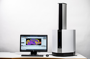
- Next Generation Mass Spectrometry Imaging by iMScope QT
- Dr Kevin Tucker, Associate Professor Southern Illinois University, Edwardsville
Easy to use MALDI-8020 imaging reveals local differences in analyte concentrations and makes this technique a cornerstone in analyte quantitation workflows - Kratos – MALDI Manufacturing and R&D Centre in Manchester
Applications Team Interviews - Benchtop Mass Spectrometry Imaging using MALDI-8020/8030
Highlights
Dr Ruth Gordillo, Southwestern Metabolic Phenotyping Core, University of Texas
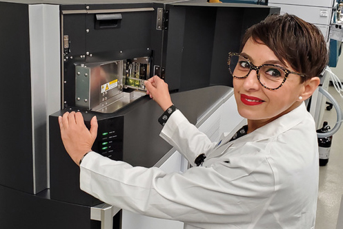
Dr Ruth Gordillo was born in Badajoz, Spain. She earned her B.S. and Ph.D. degrees in Chemistry from the University of Extremadura. She came to the USA as a postdoctoral researcher at the University of California in Los Angeles in the Department of Chemistry and Biochemistry. Dr Gordillo joined UT Southwestern Touchstone Diabetes Center faculty in 2011 and became the director of the Metabolic Phenotyping core in 2015.
The Touchstone Diabetes Center is devoted to the study of the cells and tissues that contribute to or are affected by diabetes and its comorbid diseases. Research in the Touchstone Diabetes Center focuses on both basic and clinical aspects of Type 1 and Type 2 diabetes, and questions related to the impact of diabetes and obesity on cardiovascular disease, renal disease, and cancer progression.
Dr Gordillo specializes in the development of LC-MS/MS technologies for the determination and quantitation of metabolites. Our analytical portfolio includes the exploration of sphingolipids and phospholipids, water soluble cellular metabolites, diastereomeric separations of carbohydrates and nucleotide sugars, and therapeutic antibodies.
Summary of my work
I am the director of UT Southwestern Metabolic Phenotyping Core and Dr Syann Lee is the co-director. Together we manage the facility operations which provides analytical and phenotypical measures to the scientific community, both inside and outside UT Southwestern Medical Center. Our portfolio of services includes chemistry clinical metabolic panel using a clinical analyzer, colorimetric assays immunoassays, targeted metabolomics using LC-MS/ MS technologies, mice body composition (TD-NMR and micro-CT scanning), rodent metabolic chambers, rodent treadmills, mouse surgeries, and RNAscope and Base Scope hybridization.
Our goal is to expand the scope of techniques available to investigators, standardize key methodologies, and expedite the completion of research on diseases related to metabolic disorders (specifically diabetes and obesity), cancer, aging neurological disorders, etc. Amongst a large portfolio of phenotyping techniques, our laboratory allocates extensive mass spectrometry resources to metabolomics studies.
I specialize in the development and implementation LC-MS/MS methodologies for the determination and quantitation of metabolites. Our analytical portfolio includes the exploration of sphingolipids and phospholipids, water soluble cellular metabolites, diastereomeric separations of carbohydrates and nucleotide sugars, and therapeutic antibodies.
Instrumentation and applications
Amongst a large portfolio of phenotyping techniques, our laboratory allocates extensive mass spectrometry resources to metabolomics studies.
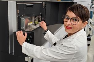
We have several Shimadzu liquid chromatography triple quadrupole mass spectrometry systems dedicated to the analysis of lipids and water-soluble primary metabolites. A supercritical fluid chromatographer coupled to a LCMS-8060 for more challenging separations such as diastereomeric glycosphingolipids and phosphatidic acids. In addition, we have allocated an LC/Q-TOF-MS dedicated to the analysis of free fatty acids profiling and global lipidomics studies. A fully automated sample preparation module coupled to a LCMS- 8060 LC-MS/MS system. An important percentage of our data is obtained using the ready to use method packages developed by Shimadzu such as the library for phospholipid profiling, cell culture profiling, primary metabolites, and bile acids quantitation. Very recently we have installed a mass spectrometry imaging system, a Shimadzu iMScope QT, expanding our mass spectrometry resources dedicated to metabolomics. Imaging mass spectrometry is a very necessary and logical step in our laboratories to be able to answer many questions related to our studies on preclinical models of disease. When we perform LC-MS/MS metabolite analysis on tissue extracts we lose the spatial resolution and subtle spatially localized metabolite differences are also lost. We are currently embarked on the method implementation and optimization processes for MS lipidomics and bile acids imaging. Our immediate plans include studies focusing on liver, kidney, and pancreas.
In the future we hope to expand the applications to proteomics imaging and therapeutic drugs in collaboration with UTSW Proteomics Core and the Preclinical Pharmacology Core. iMLayer sublimation and iMLayer AERO automatic sprayer satellite instrumentation have been very powerful tools in our method implementation journey assisting with sample preparation and reproducibility. Having a straightforward and reproducible processes is of utmost importance in our facility since we provide services to the scientific community and many of the steps are performed by different research technicians.
What do I like about working with Shimadzu?
There are many aspects I like regarding working with Shimadzu. Their extensive catalog of analytical instrumentation allows us to have a large number of fully compatible systems in our laboratories. They have a modular design concept facilitating laboratory expansions and rearrangements. Shimadzu mass spectrometers have ultra-fast-scanning capability, are very sensitive and very robust. We have been able to apply our analytical methods to all kind of complex biological matrices as well as perform metabolomic studies on samples with a very small amount of biological material such as mouse and human pancreatic islets preparations and small extracellular vesicles preparations. But what I appreciate the most about collaborating with Shimadzu is the support I get from their team of engineers and scientists. I am extremely fortunate to work with very talented professionals from Japan, Europe, and the USA.
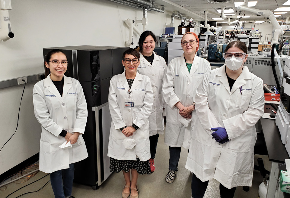
Image Dr Gordillo with her team from left to right: Jannine Irel Gamayot, Ruth Gordillo, Sarah Rico, Shannon Hacker, Allison Walker
Dr Kevin Tucker, Associate Professor, Southern Illinios University, Edwardsville
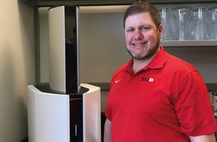
Dr Kevin Tucker, Associate Professor at Southern Illinois University Edwardsville, tells his story of using complementary mass spectrometry analyses to understand localization and concentration of analytes in a variety of matrices.
Summary of my work
Summary of my work In 2005 when I started at the University of Illinois at Urbana- Champaign, I became enamored with the future possibilities of mass spectrometry imaging (MSI) even though at the time MSI was still a burgeoning technology. While studying under Prof. Jonathan Sweedler, I grew as a mass spectrometrist and an analytical chemist, focusing my time and skills on method development specifically for MSI based techniques. My time spent in the lab and my dedication to bolstering my MSI knowledge led me to becoming proficient in improvising techniques and improving data quality for MSI. After matriculating from my Ph.D. in 2011, I found myself performing qualitative and quantitative analyses on a variety of samples that spanned the gamut from nucleotides to ground corn to concrete. I had become adept at method development for a very broad range of applications. My independent research career began in 2016 and has focused on the application of method development for LC-MS and MALDI MSI to a variety of sample types and sample matrices. Utilizing an LC QqQ MS, I have performed targeted quantitative analysis on samples including wastewater, corn-to-ethanol fermentation broth, and homogenate tissue samples. However, the most exciting work has come from marrying my two areas of expertise: LC-MS quantitation method development and MALDI MSI method development.
Instrumentation and applications
Instrumentation and applications I lead the work that is performed at Southern Illinois University Edwardsville’s Shimadzu Innovation Lab, which currently houses a HPLC iSeries, a LCMS-2020, a LCMS- 8060NX, a GCMS-TQ8050, a MALDI-8020, an EDX-8100, and a UV/Vis 3600i. I have been able to leverage these analytical tools against a plethora of applications: antibiotics in freshwater, wastewater, soil, and dried distiller’s grains with solubles; pesticides and herbicides in fresh water, plants, and foods; pharmaceutical toxicology in flatworms, fathead minnows, leeches, and earthworms; targeted metabolomics in gynecological cancers and blood; and the application of qualitative MALDI MSI whenever appropriate to guide the quantitative studies. The recent addition of the MALDI-8020 has been remarkable as it allows me to train undergraduate and graduate students with little to no experience with MSI and produce publication quality data within the first couple of months. My students found the software to be easy to follow with very little training required. As a result, I have had three students using the instrument consistently for imaging work related to earthworms, human cancer, and mung beans. We have employed the MALDI MSI to inform our studies on where the analytes of interest are most concentrated in the tissue. We then perform careful dissections of the samples to maximize the concentrations of our analytes. Performing MALDI MSI is now the cornerstone in our workflow to quantifying those analytes that are present at high local concentrations, but low global concentrations and are thus diluted in homogenate sampling protocols. What do you like about working with Shimadzu?
What do I like about working with Shimadzu?
In addition to Shimadzu’s excellent instruments, their work force is what sets their organization apart from other manufacturers. I know that I can get everything I need out of my instruments because I have direct contact with the experts at Shimadzu anytime I need help. Whether by email or phone, Shimadzu’s employees have not only provided the tools that researchers need on their journey to excellence in science, but are willing to walk beside you to help you along the way as you encounter new challenges that even they may not be familiar with yet. Working with Shimadzu for me means that I am working people who are invested in my success.
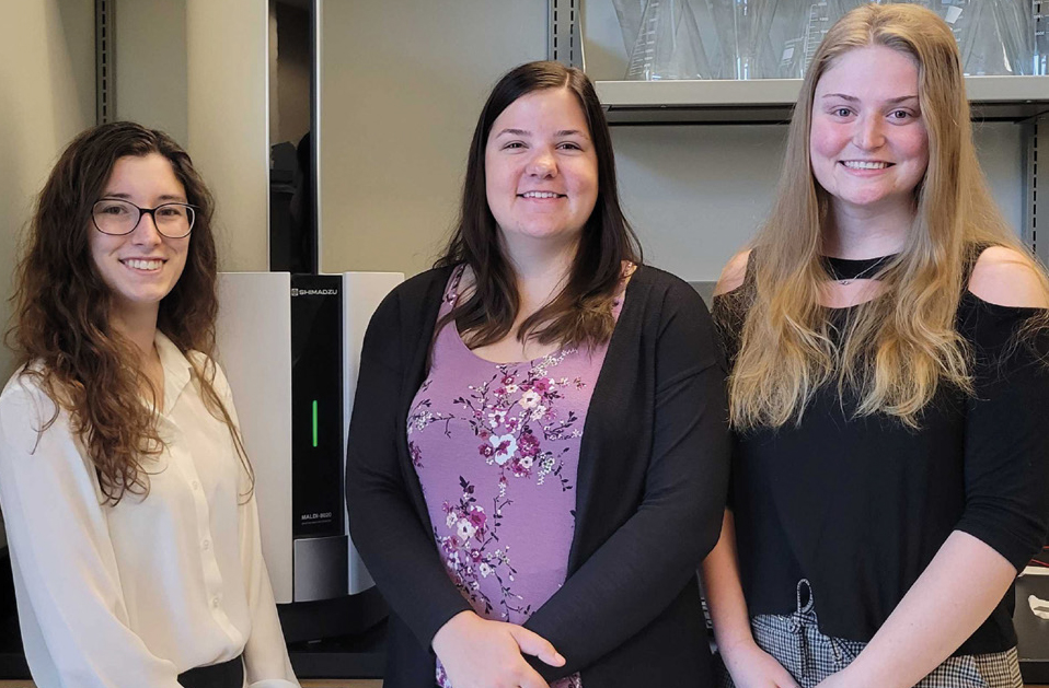
Images Dr Kevin Tucker and his students next to their MALDI-8020 at their lab at SIUE (Southern Illinois University Edwardsville). His grad students are (left-to-right): Kendra Selby, Emily Hubecky and Stephanie Shan


